Last updated on May 15th, 2022
The human heart is a muscular organ; it pumps blood through the blood vessels. It is the size of a fist (about 13 centimeters long and 9 centimeters broad) and is located in the middle compartment of the chest, behind and slightly to the left of your breastbone. With these facts about heart, let us uncover more about its anatomy, working, relationship with other body parts and more…
About its anatomy
1. There are four chambers (each chamber holds about 70 ml of blood) in the human heart. The total volume of the heart is therefore approximately 4 times that value, or 280 ml. There is upper right and left atria and there is lower right and left ventricle. Each of these chambers has a one-way valve which is positioned at its exit. These valves prevent blood from flowing backward.
2. The heart can be divided into two parts; the left and the right heart. The right heart includes the right atrium and the right ventricle while the left heart includes the left atrium and the left ventricle.
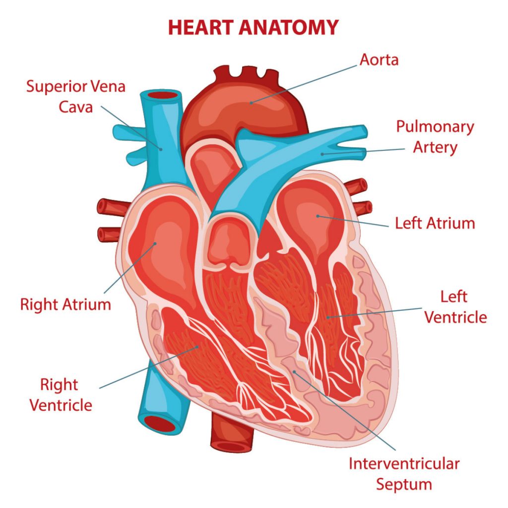
3. The blood inside of the heart flows only in one direction. The four valves (tricuspid, pulmonary, mitral and aortic) in the heart help keep the blood flow in check. The blood enters the heart through the right atrium and then moves to the right ventricle where it is pumped to the lungs. And after picking up oxygen from the lungs, it moves to the left atrium and then leaves it for the left ventricle where it is finally supplied to the body. The heart pumps out about 70 ml of blood with each beat. [Note: an average adult body with a weight of 150 to 180 pounds will contain approximately 4.7 to 5.5 liters (1.2 to 1.5 gallons) of blood.]
4. There are three layers of the heart wall: Epicardium (the outermost layer), myocardium (the muscular middle layer) and endocardium (the inner layer). The function of Epicardium is to protect the inner layers and to assist in the production of pericardial fluid.
5. The two sides of a human heart are separated by a septum, which is essentially a wall of muscles in the heart (see the anatomy of the heart above.)
6. Atrias are smaller than the ventricles, and their walls are thinner. The job of the ventricles is to pump blood. The right ventricle pumps blood to the lungs while the left ventricle pumps blood to all other parts of the body. Note that the wall of the left ventricle is stronger than the wall of the right ventricle. It is also a fact that the left ventricle is the strongest of all the four chambers of the heart.
7. The Superior vena cava carries blood from upper body parts such as head, neck and upper limbs to the heart while the inferior vena cava carries blood from the other body parts to the heart.
8. The pericardial cavity is the place where the heart sits. It is a fluid-filled cavity with walls and linings made from a special membrane known as the pericardium. The purpose of the fluid is to lubricate the heart and prevent the friction between it and its surroundings.
9. The heart’s weight is less than 0.5 percent of the total body weight of a person.
About heart’s functioning
10. Did you know that each heartbeat fills all the four chambers of the heart with a fresh round of blood?
11. The heart lies at the center of the blood delivery system. It is an essential part of the entire system, which when becomes dysfunctional can lead to death. The heart pumps oxygen and nutrient-rich blood (blood is composed of cells and plasma) to organs, tissues, and cells of your body. Blood also serves an important function of removing carbon dioxide and waste products made by those cells.
12. The heart also has plenty of pacemaking cells that determine the blood flow. Every pacemaking cell can become a “band leader” and the rest of the cells will follow that cell. However, when many of these cells become bandleaders, they fall out of rhythm and the heartbeat becomes irregular, which is usually a concern for the person.
13. The heart is made of cardiac muscles that work involuntarily. The heart beats automatically in response to the nerve signal from the brain.
14. The superior vena cava and the inferior vena cava are the two largest veins that carry blood into the heart.
15. The heart receives blood which is low in oxygen and then the blood passes through the lungs where it is oxygenated. This oxygen-rich blood again enters the heart and is then sent to the body.
16. The resting heart rate in adults is between 70 – 72 beats per minutes. Check the data for heart rate according to the age of an individual: Newborn: 130 bpm; 3 months: 140 bpm; 6 months: 130 bpm; 1 year: 120 bpm; 2 years: 115 bpm; 3 years: 100 bpm; 4 years: 100 bpm; 6 years: 100 bpm; 8 years: 90 bpm; 12 years: 85 bpm; adult: 60 – 100 bpm.
17. The heart is placed between the lungs. Thus, the left lung is usually smaller than the right lung to accommodate the heart.
18. Did you know that a lower resting heart rate indicates a high level of cardiovascular fitness? The resting heart rate of athletes could be as low as 40 beats per minute.
19. Stress and anxiety is the most common cause for a fast heart rate (also known as pulse).
20. Well trained athletes have a larger heart because it has to pump more blood.
21. Did you know that the heart is the first functional organ to develop and start to beat and pump at about 3 weeks into embryogenesis?
22. The heart receives nerve signals from the vagus nerve and from the nerves arising from the sympathetic trunk.
23. The function of vagus nerves (also called X cranial nerve or 10th cranial nerve, longest and most complex of the cranial nerves) is to decrease the heart rate while that of the sympathetic trunk is to increase the heart rate. Vagus nerve is the longest nerve of the autonomic nervous system in the human body.
24. Did you know that the vigorous contraction of our left ventricle creates our blood pressure? The blood pressure in the heart is the greatest in the left ventricle. Because this is the place where the blood leaves the heart and for it to flow throughout the system, it has to have a high pressure.

25. We have by far known that the heart’s function is to keep the cells in the body energized by supplying them with oxygenated blood. But did you wonder from where does the heart receiver power for its own functioning? The heart receives its power through the coronary arteries that run along its surface and supply it with oxygen-rich blood. Thus, it keeps functioning and keeps pumping blood for other body parts. The heart utilizes 4-5% of its blood output through the coronary arteries for its own functioning.
26. Blockage in the coronary arteries causes the heart attack.
27. Blood vessels include arteries, veins, and capillaries. The function of arteries is to take the blood away from the heart while that of the veins is to bring the blood back to the heart.
28. If you have ever heard the sound of a functioning heart, you would have heard “lub-dub”. This is the typical sound of a properly functioning heart. The sound is actually produced due to the shutting of the valve on the blood. The function of the valves is to prevent the backflow of the blood.
29. When we run, our body needs more oxygen to supply to the cells for exaggerated physical activity. In this scenario, our heart beats faster and delivers the much-needed oxygen for the activity.
30. Your heart is a muscular pump. Regular exercising is beneficial for your hearts health.
31. Note that a cardiac arrest is different from a heart attack. In case of a cardiac arrest, a sudden loss of heart function happens due to loss of electrical sensation to the heart. Moreover, a heart attack can lead to a cardiac arrest.
32. The normal rhythmic heart rate is called “Sinus Rhythm”. It is created by the Sinoatrial Node. With the help of specialized fibers, electrical impulses are conducted from the Sinoatrial node to the rest of the heart.
33. The electrical impulse that drives the heart is created at the Sinoatrial node located at the upper part of the right atrium near to the junction with superior vena cava. Pacemaker cells are responsible for producing electrical impulses in the heart. These cells are like the driving force which keeps our heart in momentum. There are plenty of these cells in the heart.
34. Our heart does not rest at all. Even when you are sleeping, it is still functioning. It does not get any day off from its work.
35. Interestingly, the heart plays a significant role in our mental, emotional and physical processes.
36. In humans, the heart rate at the time of the birth is 130 bpm and that for an adult is 70-72 bpm. However, in the old age, the heart rate increases slightly.
37. Note that the heart rate during sleep decreases.
38. Unfortunately, the heart diseases are a leading cause of death in UK, USA, Canada, and Australia. Cardiology is the study of the human heart and its various diseases.
39. The heart goes through many developmental stages. In the fetus, it is first similar to the heart of a fish, and then it becomes similar to that of a frog with two chambers, and in a later stage, it turns like that of a snake or turtle heart which has three chambers. And finally it takes the shape of a human heart with four chambers.
40. Did you know that a special stethoscope called a fetoscope is used by the doctors to listen to the fetal heartbeat? The presence of a fetal heartbeat confirms pregnancy. The fetus has a much faster heart rate than the mother. Moreover, it is not so easy to distinguish between the mother’s and fetal heartbeat.
41. Aerobic exercises such as walking, swimming, and biking are good for the heart’s health. Because the heart is a muscular thing, exercising naturally makes it stronger and healthier. Pay attention that people who don’t exercise are almost twice as likely to get heart disease as people who are active.
Interesting facts
42. Did you know that generally, the rate of heartbeat varies inversely with the size of the animal? Learn that the heart rate of an elephant (the largest land animal and the longest-lived animal as well – body mass averages between 2300 to 7000 kilograms) is 25 beats per minute while that of a canary (body mass is 20 grams, approximately) is 1000 beats per minute. The heart rates of other animals are rat – 420 bpm, horse – 38 bpm, rabbit – 205 bpm, and a large dog – 85 bpm. The heart rates of some of the birds are pigeon – 185 bpm, hen – 312 bpm, turkey – 193 bpm.
43. The heart generates a powerful electromagnetic field. When the electromagnetic field of the heart was measured on an electrocardiogram (ECG), it was found that it was about 60 times greater in amplitude than the brain waves recorded in an electroencephalogram (EEC).
44. The electromagnetic field of the heart is so powerful that it can be measured several feet away from an individual’s body.
45. Did you know that when a heart transplant is made, the doctors have a narrow window of 4-6 hours to replant the removed heart in the receiver before it becomes unusable?
46. The human heart pumps blood through a 60,000 miles long network of vessels (arteries, arterioles, capillaries, venules, and veins.)
47. Did you know that it is possible to live without a real heart? Yes, a 25-year-old Ypsilanti, Michigan, resident lived a whopping 555 days before he received a real heart from a donor for his heart transplant. Unfortunately twenty-two Americans die waiting for heart transplants daily.
48. Some studies also relate baldness to a much greater risk of coronary heart disease.
49. Interestingly, the human heartbeat changes and mimics the music you listen to.
50. Interestingly, sex is good for a healthy heart. And research suggests that it can bring down the chances of a heart attack by fifty percent.
51. The blood pressure in the left ventricle of the heart is enough to squirt the blood to a height of 30 feet in the open air.
52. There is blood supply to every part of the human body except the corneas. Also, note that the heart supplies blood to almost 75 trillion cells in the human body. Astonishing, isn’t it!
53. The Stethoscope was invented because it was inappropriate for the doctor to place his ear on his female patients’ chest to hear the heart sound.
. . . continue reading on the next page.